[et_pb_section][et_pb_row][et_pb_column type=”4_4″][et_pb_text admin_label=”Text” background_layout=”light” text_orientation=”left” use_border_color=”off” border_color=”#ffffff” border_style=”solid”]
The Case:
A 27 year old man with a past medical history of Crohn’s disease requiring three bowel resections presented with two days of generalized non-radiating abdominal pain associated with 10 episodes of brown colored emesis, lack of bowel movements, and decreased oral intake. On physical exam, he was noted to have diffuse abdominal tenderness, abdominal distention. A CT scan was ordered for evaluation of the abdomen, and bedside sonogram of the right upper quadrant was requested to evaluate for cholelithiasis or free fluid. He has no known history of SBO (Small Bowel Obstruction).
The following images were obtained (click on arrows or on image for gallery):
[/et_pb_text][et_pb_gallery admin_label=”Gallery” gallery_ids=”1787,1786,1788″ fullwidth=”on” show_title_and_caption=”on” show_pagination=”on” background_layout=”light” auto=”off” hover_overlay_color=”rgba(255,255,255,0.9)” caption_all_caps=”off” use_border_color=”off” border_color=”#ffffff” border_style=”solid”] [/et_pb_gallery][et_pb_text admin_label=”Text” background_layout=”light” text_orientation=”left” use_border_color=”off” border_color=”#ffffff” border_style=”solid”]
The first two images are of the abdomen with dilated loops of bowel clearly visualized within peritoneal free fluid. The last image shows a fluid filled and distended stomach visualized in the left upper quadrant when attempting to evaluate the splenorenal recess for free fluid.
[/et_pb_text][et_pb_divider admin_label=”Divider” color=”#ffffff” show_divider=”off” height=”20″ divider_style=”solid” divider_position=”top” hide_on_mobile=”on”] [/et_pb_divider][et_pb_text admin_label=”Text” background_layout=”light” text_orientation=”left” use_border_color=”off” border_color=”#ffffff” border_style=”solid”]
A CT scan of the abdomen and pelvis was subsequently performed (click on arrows or on image for gallery):
[/et_pb_text][et_pb_gallery admin_label=”Gallery” gallery_ids=”1794,1795,1793,1796″ fullwidth=”on” show_title_and_caption=”on” show_pagination=”on” background_layout=”light” auto=”off” hover_overlay_color=”rgba(255,255,255,0.9)” caption_all_caps=”off” use_border_color=”off” border_color=”#ffffff” border_style=”solid”] [/et_pb_gallery][et_pb_text admin_label=”Text” background_layout=”light” text_orientation=”left” use_border_color=”off” border_color=”#ffffff” border_style=”solid”]
The Outcome
CT was read as small bowel obstruction with transition point at the ileocolic anastomosis. Abdominal X-ray was not performed on this patient as part of his initial workup. Ultimately, this patient was admitted to the surgical service and managed conservatively with a nasogastric tube.
The Discussion
Several imaging modalities are available for evaluating post-surgical patients with abdominal pain, including abdominal X-Ray, abdominal CT, and ultrasonography. Point of Care Ultrasonography is gaining in popularity for initial diagnosis or evaluation of patients with abdominal pain thought to be secondary to SBO. Two prospective ED based studies have been performed to evaluate the diagnostic accuracy of point of care ultrasound for SBO.1-2 One enrolled 76 patients and compared abdominal X-Ray with bedside ultrasonography in the ED for detecting small bowel obstruction (SBO); CT was the “gold standard” in this study.1 The researchers found that dilated bowel on US was 91% sensitive and 84% specific for diagnosis of SBO, compared to 41.2% sensitivity and 66.7% specificity on abdominal X-ray when the read was diagnostic. The other enrolled 174 patients.2 They found a sensitivity of 98% and a specificity of 93% when compared to CT or surgical findings.
The Procedure
- Grab the curvilinear probe
- Mow the Lawn – Evaluate each area of the abdomen successively, looking for an area of obstruction, as seen in the following image. While various techniques exist we feel this is the most comprehensive.
- Look for suspicious findings:
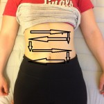
Scanning technique. Posted with permission. - Dilated fluid filled loops: The most important sonographic finding. Fluid filled bowel loops >3cm in diameter adjacent to collapsed bowel.
- Thickened bowel wall (>3mm), and increased motion of bowel contents.
- Bowel wall edema and free fluid in the abdomen indicate a higher grade of obstruction and may be a sign of impending bowel strangulation and ischemia.
- Gastric distention may also be noted.
- Transition points may be visualized on ultrasound, but are difficult to assess.
Our Conclusion
Current studies indicate that – in the right hands – bedside US is comparable to CT scan in diagnosis of small bowel obstruction. This is particularly important in the ED setting where a more rapid diagnosis of SBO could lead to earlier intervention and better patient outcomes. The same is true of limited resource and remote/wilderness settings. Serial US exams during hospitalization may also help evaluate the progression of SBO in patients undergoing non-operative management. However, the studies to date have been small and do not reflect general practice in large academic centers where the patient population may not be comparable and a wide range of providers at different levels of medical training may be involved in the patient’s care and diagnosis. Larger studies at a greater variety of medical centers would be helpful to further support the use of bedside US in evaluation of patients with suspected SBO.
[/et_pb_text][et_pb_divider admin_label=”Divider” color=”#ffffff” show_divider=”off” height=”30″ divider_style=”solid” divider_position=”top” hide_on_mobile=”on”] [/et_pb_divider][et_pb_text admin_label=”Text” background_layout=”light” text_orientation=”left” use_border_color=”off” border_color=”#ffffff” border_style=”solid”]
References:
- Jang TB1, Schindler D, Bedside ultrasonography for the detection of small bowel obstruction in the emergency department. Emerg Med J.2011 Aug;28(8):676-8.
- Unlüer EE1, Yavaşi O, Eroğlu O, et al. Ultrasonography by emergency medicine and radiology residents for the diagnosis of small bowel obstruction. Eur J Emerg Med.2010 Oct;17(5):260-4.
[/et_pb_text][/et_pb_column][/et_pb_row][et_pb_row][et_pb_column type=”1_2″][et_pb_team_member admin_label=”Person” saved_tabs=”all” name=”Shayna Carp, MD” position=”Author” image_url=”http://theempulse.org/wp-content/uploads/2015/11/Shayna-Carp.png” animation=”off” background_layout=”light” use_border_color=”off” border_color=”#ffffff” border_style=”solid”]
Shayna Carp is a Categorical Emergency Medicine resident at LIJ Medical Center.
[/et_pb_team_member][/et_pb_column][et_pb_column type=”1_2″][et_pb_team_member admin_label=”Person” name=”Dr. Josh Guttman, MD, FRCPC” position=”Reviewer and Ultrasound Editor – theEMPulse.org ” image_url=”http://theempulse.org/wp-content/uploads/2015/10/Guttman-e1453253661766.jpg” animation=”off” background_layout=”light” twitter_url=”https://twitter.com/josh_guttman” use_border_color=”off” border_color=”#ffffff” border_style=”solid” saved_tabs=”all”]
Dr. Guttman graduated from the at Mt. Sinai Medical Center Emergency Ultrasound Fellowship and is now an Attending Physician at the LIJ Division of Emergency Ultrasound.
[/et_pb_team_member][/et_pb_column][/et_pb_row][et_pb_row][et_pb_column type=”4_4″][/et_pb_column][/et_pb_row][/et_pb_section]