Case:
A 20 year old female presented to the ED after having stepped on a sewing needle 3 days earlier, having it now buried in the plantar surface of her left foot. She described pain when she walked, but no fevers, chills or other systemic symptoms. Her vital signs were normal and she was well appearing. The plantar surface of her foot revealed a small area of swelling and erythema. There was no visible or palpable foreign body. An xray was done, revealing the needle. In order to facilitate removal of the needle, an ultrasound guided posterior tibial nerve block at the ankle was performed, rendering complete analgesia of the plantar surface of the foot. After exploration, a 1 cm pin was extracted. The patient remained pain free and comfortable, playing on her phone throughout the procedure.
Discussion:
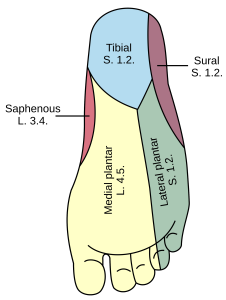
Lacerations and foreign bodies to the sole of the foot are common presentations to the ED. Adequate local anesthesia is difficult and painful due to rough skin of that area. Local anesthesia to majority of the sole of the foot can be adequately achieved with a posterior tibial nerve block. While the sural nerve does innervate a small part of the plantar surface, the vast majority of lacerations and foreign bodies are located in the distribution of the posterior tibial nerve. This single block is therefore enough for ED procedures on the sole of the foot. In the image to the right, the Tibial, Medial Plantar and Lateral Plantar nerves are all branches of the posterior tibial that will be anesthetized by a successful PT Block.
Performing a PT Block:
1)Assemble your equipment
- Long 20-25 gauge needle
- 10 cc syringe with Lidocaine 2%, with or without epinephrine
- Ultrasound machine with linear probe
- Sterile ultrasound gel, such as surgilube
- Tegaderm or probe cover
- Chloroprep or other antiseptic swab
2) Place the ultrasound probe just posterior to the medial malleolus
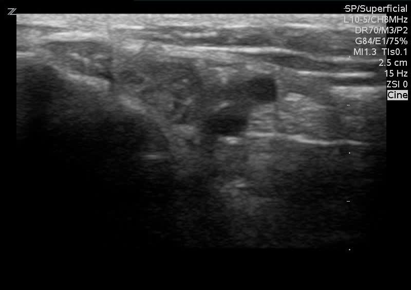
3)Identify the nerve in cross-section. Start by identifying the posterior tibial artery, which should be pulsating and easy to identify. Use color if needed. The nerve is located just posterior to it. If unsure where the nerve is, follow the artery proximally until the nerve is easily identified. Note that in this image the nerve is just adjacent to the artery. Therefore you would approach the nerve from the other side of the probe when performing the block.
4)Insert the needle in long axis
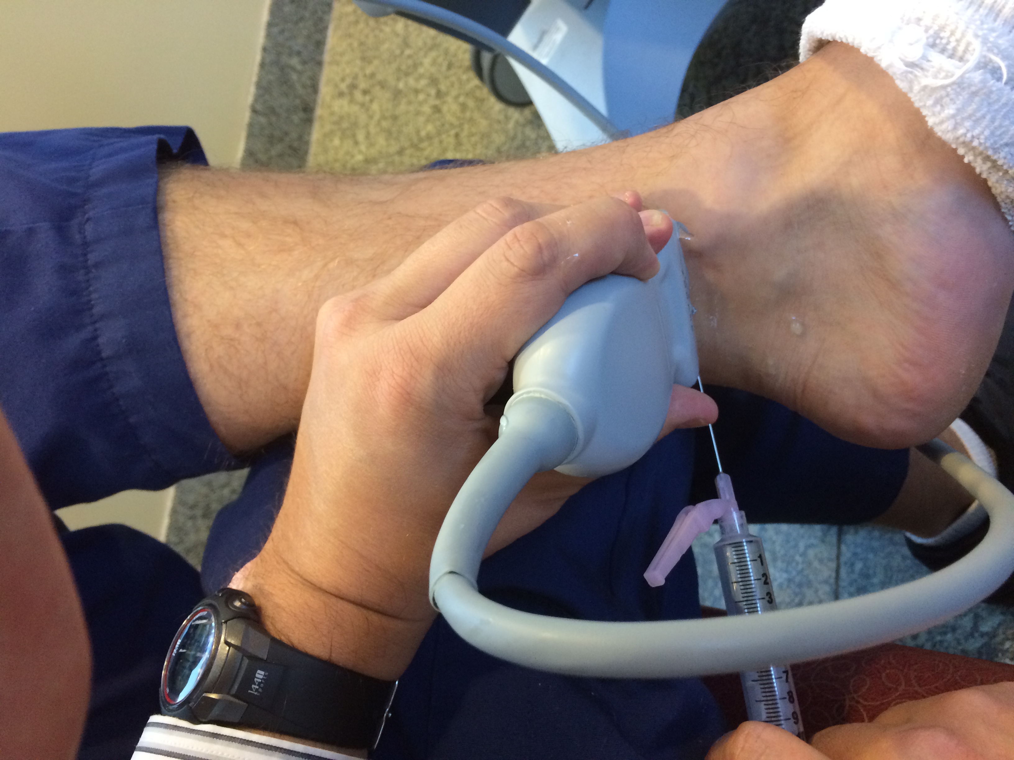
5)Follow the needle tip to the nerve, without hitting the nerve
6)Inject lidocaine and watch the anechoic fluids spread. Adjust the needle depth to inject lidocaine below and above the nerve.
Tips and Tricks:
- As in any ultrasound procedure, make sure to “pre-scan” to makes sure you can identify the nerve in the appropriate location
- When scanning, first identify the posterior tibial artery, which is easy to see because it pulsates. The nerve is located just posterior to it. This will be either to your right or left on the machine, depending on which direction the probe marker is.
- While following the needle, make sure to fan the probe back and forth, to make sure you are seeing the entire length of the needle.
- If you lose site of the needle tip, aspirate, then inject a little lidocaine. This is termed “hydrodissection” and the anechoic fluid will allow you to have an easier time identifying the needle.
- As in any ultrasound procedure, if you don’t know where you are, STOP, pull the needle out and start again.
Thanks to Dr. Ilya Parizh for photographic assistance