Case:
A 50 year old female with a history of only iron deficiency anemia presented to the ED with 5 hours of a gradual onset of epigastric pain radiating into the chest as well as nausea, associated with one episode of vomiting. She had never experienced similar pain. Her ECG was normal and her lab work revealed a WBC count of 13.3, normal LFTs, Lipase and troponin levels. Her CXR was normal.
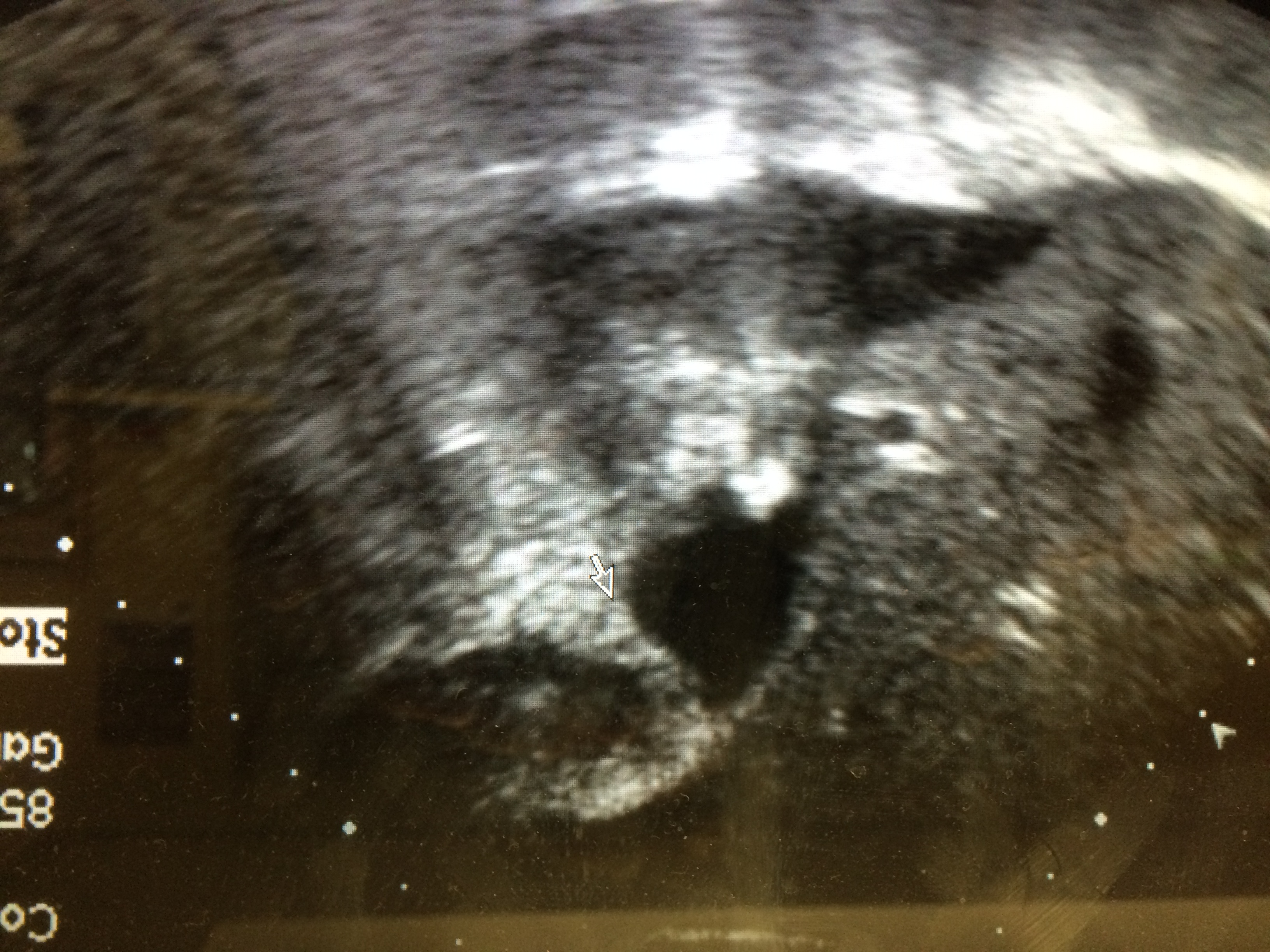
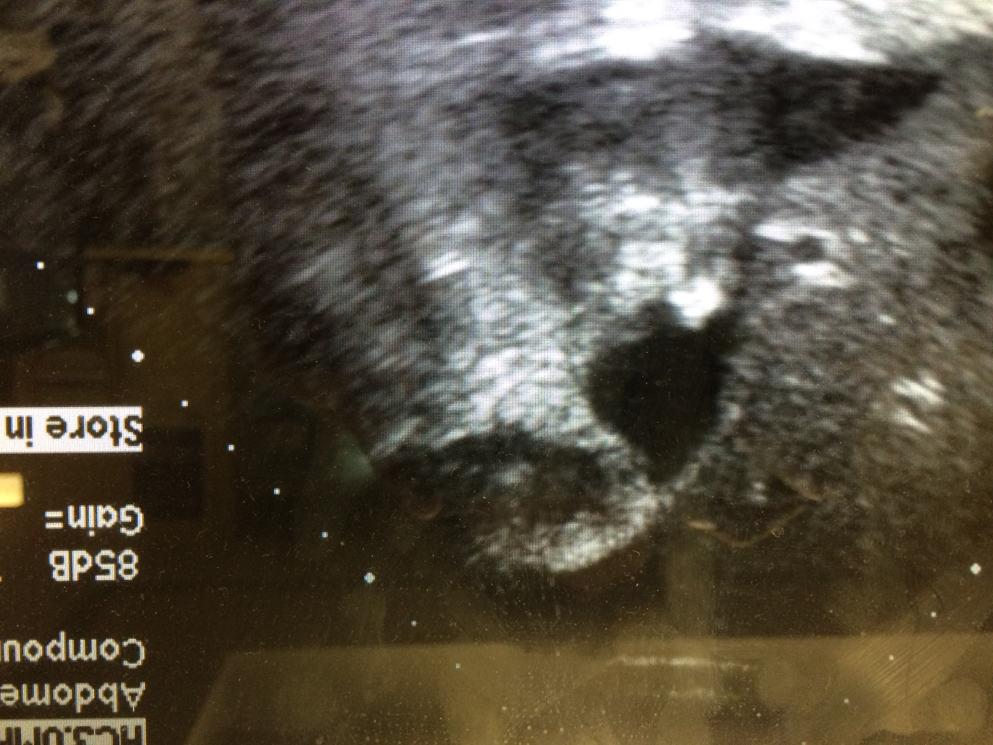
A bedside ultrasound was initially read by the resident as gallstones without any evidence of cholecystitis. A repeat bedside ultrasound noted gallbladder wall thickening localized to the gallbladder fundus of 5.7 mm with some associated pericholecystic fluid. Generally surgery was then consulted, who requested a radiology ultrasound revealing similar findings,
The patient was then admitted and underwent cholecystectomy.
Discussion:
Cholecystitis is the most common cause of gallbladder wall thickening in the USA. It is, however, a non-specific sign that can be associated with other diseases, including pancreatitis, diverticulitis, hepatitis, pyelonephritis and CHF. Therefore, clinical context and associated signs must be taken into consideration. When the clinical presentation suggests cholecystitis, several ultrasound findings suggest the diagnosis. These include the findings of gallstones or sludge in the gallbladder, sonographic murphy’s sign, gallbladder wall thickening, pericholecystic fluid, gallbladder enlargement (greater than 4 cm in transverse diameter). Ultrasound has a sensitivity of 88% and specificity of 80% for acute cholecystitis.
Wall thickening associated with acute cholecystitis is generally diffuse, occupying the fundus, body and neck of the gallbladder. In patients presenting to the ED with suspected acute cholecystitis or cholelithiasis, ultrasound may reveal a focal wall thickening. While this may be seen with acute cholecystitis, it is more commonly found with chronic cholecystitis, a non-emergent disease process. Additionally, focal gallbladder wall thickening may also represent gallbladder carcinoma.
Conclusion:
While focal gallbladder wall thickening may be due to acute cholecystitis, it is more commonly associated with chronic cholecystitis. Additionally, focal gallbladder wall thickening may be associated with gallbladder cancer and therefore expedient radiology imaging and follow-up should be arranged\
References:
Barbosa ABR, Souza LRMF, Pereira RS, D’Ippolito G. Gallbladder wall thickening at ultrasonography: how to interpret it? Radiol Bras.2011 Nov/Dez;44(6):381–387.
Shea JA, Berlin JA, Escarce JJ, et al. Revised estimates of diagnostic test sensitivity and specificity in suspected biliary tract disease. Arch Intern Med. 1994;154(22):2573
Kim BS, Oh JY, Nam KJ et al. Focal thickening at the fundus of the gallbladder: computed tomography differentiation of fundal typeadenomyomatosis and localized chronic cholecystitis. Gut Liver. 2014 Mar;8(2):219-23